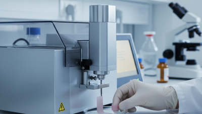
How to Use Leica Ultracut UCT Ultramicrotome Effectively?
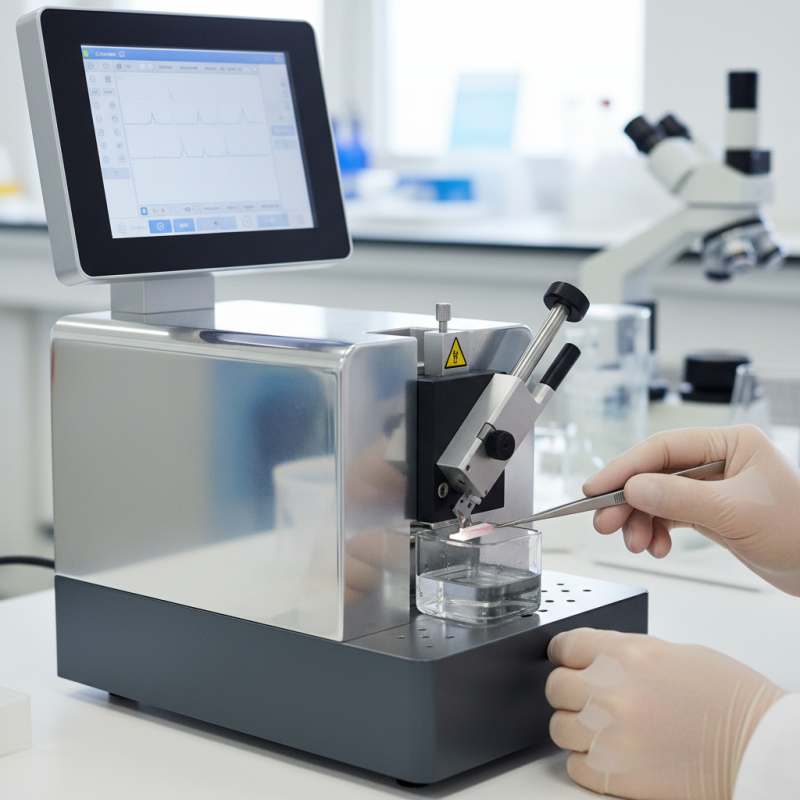
The Leica Ultracut UCT ultramicrotome is a powerful tool in histology and materials science. Its precision can create ultra-thin sections for detailed analysis. However, using this equipment effectively requires understanding its features and settings.
Many users struggle with initial setups. Calibration is crucial. Taking time to adjust the knife angle can prevent poor section quality. Operators often overlook minor details, leading to frustration. Observing how others use the Leica Ultracut UCT ultramicrotome can enhance your technique.
Learning from mistakes is vital. Each section cut can provide insight. Reflecting on how adjustments affect outcomes can refine your skills. The Leica Ultracut UCT invites experimentation, pushing users to explore diverse techniques. Embracing imperfections can lead to innovative solutions in your research.
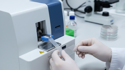
Overview of the Leica Ultracut UCT Ultramicrotome Features
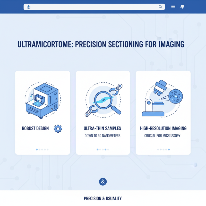
The ultramicrotome features a robust design. It focuses on precision and usability. This machine allows for ultra-thin sectioning. Users can prepare samples as thin as 30 nanometers. This capability is vital for high-resolution imaging.
Key features include an intuitive interface. The control system is user-friendly, making it accessible even for beginners. The built-in safety mechanisms enhance user confidence. However, mastering the settings takes practice. New users often struggle with the adjustments.
Another notable aspect is its temperature control. Maintaining a stable temperature is crucial during sectioning. Users sometimes overlook this, leading to sample distortion. Additionally, the blade holder is adaptable. However, it can be tricky to change blades without damaging them. Regular practice can mitigate this.
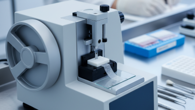
Setting Up the Ultracut UCT for Precision Cutting
Setting up the Ultracut UCT for precision cutting requires attention to detail.
The alignment of the knife is critical. A misaligned knife can result in uneven cuts, negatively affecting sample quality.
The optimal angle for the knife should be between 45 to 60 degrees. Too steep an angle can lead to blunt edges quickly.
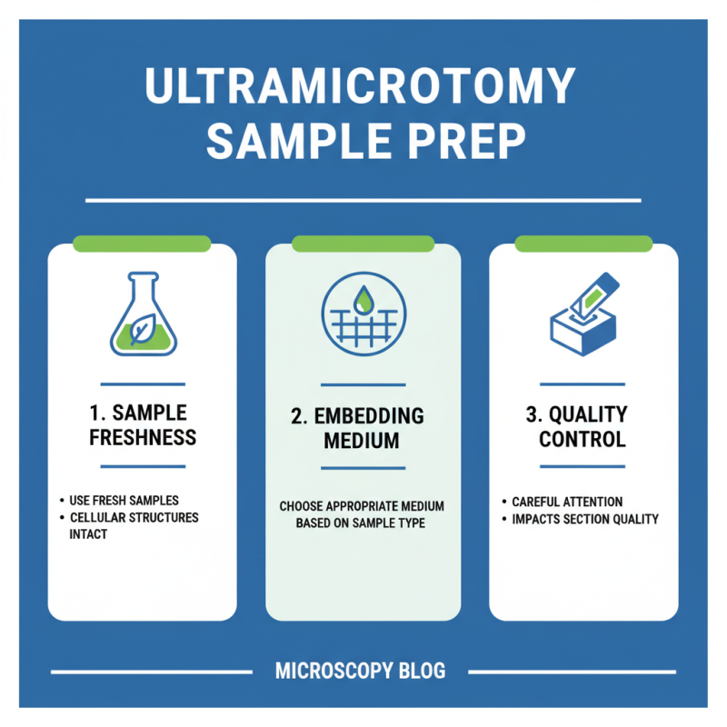
When loading samples, ensure they are properly embedded. The embedding medium should be hard enough to support thin sections.
A common issue noted in studies is the presence of compression artifacts.
Using a harder medium can reduce these artifacts, thereby improving sample clarity.
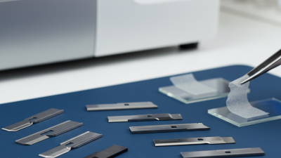
Tips: Regularly check the blade sharpness. Dull blades can produce thicker sections.
Clean the cutting area frequently to prevent debris from affecting section quality.
Also, consider environmental factors like vibration and temperature. Controlling these factors can lead to more consistent cuts.
Strive for precision, but recognize that achieving perfection takes practice. Iteration is vital for improving technique.
Techniques for Achieving High-Quality Thin Sections
Achieving high-quality thin sections is essential in microscopic studies. Many laboratories struggle with inconsistent results. Precision in sectioning is crucial. According to industry reports, inconsistent thickness can lead to misinterpretation, affecting research outcomes.
One effective technique is maintaining an optimal temperature. Keeping samples cold can improve section quality. Thin sections should ideally be between 30 to 100 nanometers. However, many users attain thicknesses of 200 nanometers or more, which complicates imaging. Such issues highlight the need for meticulous adjustments throughout the process.
Cleaning the blades regularly is another key factor. Dust or debris can create irregularities. It’s noted that using a contaminated blade can hinder the slicing process, leading to uneven cuts. Remember that even slight imperfections may alter the microscopic field of view. Awareness of these details makes a significant difference in results.
Maintenance and Troubleshooting Tips for Ultramicrotome Users
Proper maintenance of an ultramicrotome is crucial for optimal performance. Regular inspections can prevent significant issues later on. According to industry reports, 80% of equipment failures are due to inadequate care. Users should clean the cutting knife and chamber daily. Residue buildup can affect the sectioning quality. A simple wipe can save time and enhance results.
Troubleshooting common problems is essential. If sections are not uniform, check blade sharpness. Dull blades can lead to tissue compression and uneven cuts. Keep spare blades on hand for quick replacements. Adjusting the feed rate can also help. If sections stick, consider the temperature and humidity of your workspace. These factors greatly influence the cutting process.
Ultramicrotome users often face challenges. Calibration errors can lead to inconsistent results. Check alignment and settings regularly. Reports show that improper calibration accounts for 25% of user complaints. It’s vital to document calibration checks. This can reveal patterns and underlying issues. A systematic approach to maintenance not only extends equipment life but also ensures quality outcomes.