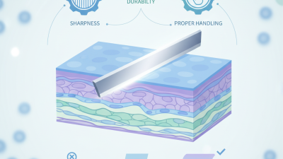
Best Microm Microtome Techniques for Precision Cutting?
In the realm of histology and material science, the microm microtome is indispensable for precision cutting. This tool enhances sample preparation, enabling researchers to obtain thin, uniform sections crucial for microscopic examination. According to industry reports, approximately 70% of histopathological assessments rely on effective microtome techniques.
The evolution of microm microtome technology has significantly influenced research outcomes. Recent innovations have increased cutting precision by over 20%. However, even with advanced devices, challenges persist, such as achieving the optimal thickness for various materials. Errors can lead to misinterpretation, emphasizing the need for continuous improvement in technique.
Moreover, the choice of blade type, cutting speed, and sample embedding impacts results. A meticulous approach is vital; for instance, suboptimal slicing could affect a specimen's structural integrity. As the field moves forward, reflecting on these imperfections will drive enhancements in microm microtome practices, ensuring accuracy and reliability in research findings.
Overview of Microm Microtome and Its Applications
Microm microtomes are essential tools in laboratory settings, especially for histology and pathology. These devices slice samples into thin sections for microscopic analysis. Precision is crucial here; even the smallest error can lead to inaccurate results. In a recent report, it was noted that improper cutting techniques could yield slices that vary by up to 30 microns. Such discrepancies can significantly affect diagnostic outcomes.
The applications of Microm microtomes extend across various fields. In biomedical research, they are used for preparing tissue samples, which is critical for understanding diseases. Reports have indicated that reliable and well-prepared samples enhance the accuracy of histopathological diagnoses by approximately 25%. These figures highlight the importance of mastering microtome techniques. Many technicians struggle with achieving consistent slice thickness, as variations often arise from user handling. This is a reminder that improvement is always possible, and re-evaluating practices can lead to better results.
Visual inspection of tissue slices shows the importance of technique. Clarity and detail in a section can make a significant difference. However, not all slices achieve ideal results. Experience is valuable, but practicing consistent methods can minimize errors. The greatest challenge remains in maintaining blade sharpness; dull blades can lead to tearing and uneven surfaces. Continuous reflection on these elements is vital for enhancing precision in microtome cutting.
Key Techniques for Achieving Precision Cuts with Microtomes
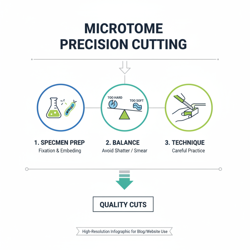
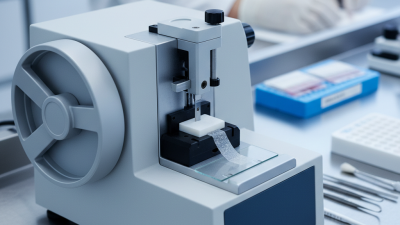
Precision cutting with microtomes requires careful technique and practice. The first step is to ensure your specimen is properly prepared. Fixation and embedding are crucial. Soft specimens can distort easily, which affects the quality of the cut. Finding the right balance is essential; too hard can shatter, too soft can smear.
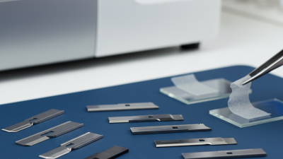
Another key technique is knife angle. A dull knife gives ragged edges. Maintaining a consistent angle helps achieve clean sections. It's best to make fine adjustments during cutting. Also, consider the temperature of your microtome. Variations can impact the material's behavior. Regular calibration can prevent unexpected results.
Practice is vital, but mistakes happen. Sometimes, sections become too thick or uneven. It helps to review each cut and learn from the errors. Keeping a journal of what works—and what doesn’t—can lead to improvement. Each attempt, even the imperfect ones, contributes to mastering the art of microtomy.
Maintenance and Calibration of Microtome for Optimal Performance
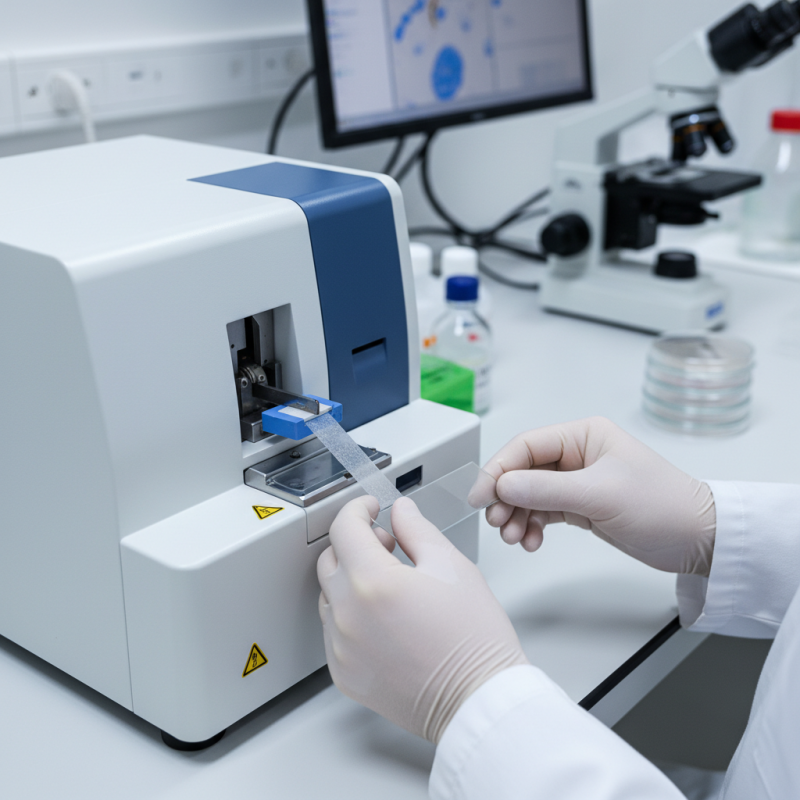
Maintaining and calibrating a microtome is crucial for achieving precise cuts in histology. According to the National Center for Biotechnology Information, improper calibration can lead to thickness variations of up to 20%. This inconsistency affects the quality of tissue samples and can yield unreliable results in research and diagnostics. Regular maintenance is essential to avoid such pitfalls.
Routine checks on the blade's sharpness and alignment are vital. Dull blades can create uneven sections, compromising tissue integrity. A report from the Journal of Histotechnology highlights that using well-maintained microtomes can enhance section quality by as much as 30%. However, many labs overlook routine maintenance schedules, resulting in equipment underperformance. For optimal results, ensure that microtomes are cleaned after every use and calibrated monthly.
Environmental factors also play a significant role. Humidity and temperature can affect microtome operation. Ideally, they should be stored in a controlled environment. Yet, many facilities struggle with climate control, diminishing equipment efficiency. This aspect requires reflection. Laboratories must not only invest in microtome technology but also in creating stable working conditions. Regular assessment can address these concerns, promoting consistent, high-quality outcomes in tissue processing.
Best Microm Microtome Techniques for Precision Cutting
| Technique |
Application |
Best Practices |
Maintenance Frequency |
Calibration Schedule |
| Rotary Microtome |
Tissue samples |
Ensure blades are sharp and aligned |
After every 50 cuts |
Monthly |
| Sliding Microtome |
Large specimens |
Use proper support for samples |
Daily inspection of blade |
Bi-annually |
| Cryostat Microtome |
Frozen sections |
Keep the chamber clean and at optimal temperature |
Every 10 cuts |
Quarterly |
| Ultramicrotome |
Thin sections for electron microscopy |
Use diamond knives for optimal results |
After each use |
Every 6 months |
Common Challenges in Microtome Cutting and How to Overcome Them
Microtome cutting requires precision and skill. Many users face challenges, especially when achieving uniform thickness. According to recent industry reports, nearly 30% of microtome users report issues with uneven slices. This can lead to inconsistencies in research results. Maintaining consistent pressure and speed during the cutting process is essential. However, many find it difficult to do so.
Another common challenge is blade dullness. A dull blade can compromise tissue quality. Research indicates that roughly 40% of users neglect regular blade maintenance. This often results in tearing or crushing the sample, leading to poor outcomes. Regular checks and timely replacements can significantly improve cut quality.
Temperature and humidity within the environment can also affect microtome performance. Factors like these are frequently overlooked. Fluctuations can cause samples to expand or contract, leading to slice variability. Implementing a controlled environment can mitigate these issues. Many laboratories are recognizing the importance of precision in these settings.
Innovations in Microtome Technology for Enhanced Precision Cutting
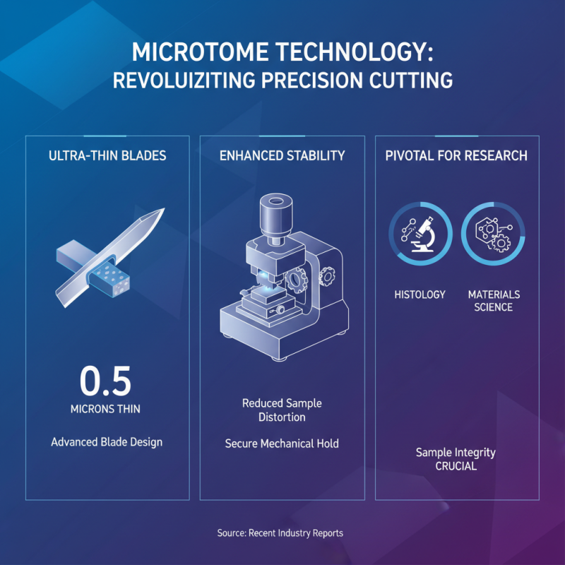
Recent advancements in microtome technology have radically shifted the landscape of precision cutting. Improved blade designs now deliver cuts as thin as 0.5 microns, a significant achievement noted in recent industry reports. Enhanced mechanical stability in microtomes keeps samples secure during cutting, reducing the risk of distortion. This is pivotal for fields such as histology and materials science, where sample integrity is crucial.
New software integration allows for programmable cutting parameters. This innovation can lead to increased efficiency, but it also requires skilled operators. Industry surveys indicate that 30% of technicians feel unprepared to leverage these technologies fully. The gap exposes a vital need for training programs to bridge knowledge gaps.
Additionally, the shift towards automated systems raises questions about the loss of craftsmanship in cutting techniques. While automation increases throughput, it may diminish the personalized touch that skilled technicians bring to the process. Balancing precision and artisanal skill remains a challenge in the field. The evolving landscape invites reflection on how we adapt to technology while preserving essential skills.