China Top Vibrating Microtome Manufacturers and Suppliers

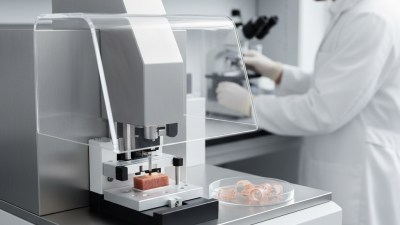
China has become an important sourcing base for laboratory sectioning equipment. Its vibrating microtome manufacturers serve universities, hospitals, pathology laboratories, and neuroscience research centers. Their products range from compact manual units to motorized systems with adjustable amplitude, speed, and blade movement.

A vibrating microtome cuts soft, unfixed, or delicate specimens without freezing them. This matters when researchers need to preserve cell layers, neural pathways, or fluorescent signals. Experienced users often notice the difference at the blade edge: clean sections float evenly, while damaged tissue curls, tears, or compresses. Small details matter.

Dr. John D. Bancroft, a respected histology author and educator, emphasized a related principle: “Good sections depend on good technique.” That observation remains useful when comparing Chinese suppliers. A machine cannot compensate for poor specimen preparation, unsuitable blades, or careless vibration settings.

Reliable manufacturers should provide clear technical data, operator training, stable after-sales support, and evidence from real laboratory use. Buyers should examine cutting thickness, specimen capacity, noise levels, safety design, and replacement-part availability. Certifications are useful, but they do not replace practical testing.

Some supplier descriptions sound impressive. Not all are equally precise. Ask for sample sections, service records, and written performance specifications before purchasing. A careful comparison can reveal whether a vibrating microtome fits routine histology, electrophysiology, botanical research, or advanced imaging work.

This guide reviews leading Chinese manufacturers and suppliers through those practical considerations. It also recognizes an uncomfortable point: the lowest price may create higher maintenance costs later. Reliability is rarely visible in a product photograph.

China Top Vibrating Microtome Manufacturers and Suppliers

Vibrating Microtomes Explained: Section Thicknesses Commonly Span 10–1,000 μm

Vibrating Microtomes Explained: Section Thicknesses Commonly Span 10–1,000 μm

A vibrating microtome can produce sections from roughly 10 to 1,000 μm thick, depending on the instrument and specimen. That range is useful, not a promise. Very thin sections may reveal fine cellular detail, while thicker slices can preserve larger structures for imaging or handling.

At the thin end, stable specimen mounting and a sharp blade matter. A slight shift can wrinkle a delicate section or make its measured thickness uneven. For thicker samples, blade clearance, vibration settings, and cutting speed influence whether the slice separates cleanly. There is no single setting that suits every tissue.

Operators often adjust the feed in small increments and inspect sections under suitable lighting. Soft tissue, dense tissue, and plant material can behave quite differently, even at the same target thickness. Temperature and preparation also affect cutting. A stated 10–1,000 μm range therefore describes broad capability; it does not guarantee equally reliable results across all samples. In practice, the best thickness is the one that preserves the feature being studied without tearing or compressing it. That judgment takes testing, and sometimes a second attempt.

China Top Vibrating Microtome Manufacturers and Suppliers - Vibrating Microtomes Explained: Section Thicknesses Commonly Span 10–1,000 μm
Topic Specification or Range Practical Context
Section thickness span Approximately 10–1,000 μm, depending on instrument and specimen This broad span describes commonly available capability; it is not a guarantee that every instrument can produce every thickness.
Thin-section planning band 10–50 μm A possible starting range for relatively thin sections; tissue condition, blade setup, and intended analysis affect the chosen setting.
Intermediate-section planning band 50–300 μm Often considered when a specimen must retain more three-dimensional structure than a thin section provides.
Thick-section planning band 300–1,000 μm Useful as a planning range for thicker slices, including preparations where maintaining larger tissue structures is important; suitability depends on the sample and downstream method.
Cutting action A blade moves with controlled lateral vibration while advancing through the specimen The vibrating action is designed to help section soft, often unfixed or lightly fixed, tissue while it is supported in a suitable medium.
Common specimen approach Fresh or fixed tissue; embedding or support methods vary by protocol Agarose support is commonly used for delicate or small specimens. The appropriate preparation should be validated for the tissue and application.
Operating conditions Blade vibration, advance rate, blade angle, and specimen temperature These settings influence section quality and should be optimized for the specimen rather than assumed to be universal.
Supplier specification checks Usable thickness range, adjustment resolution, blade compatibility, stage travel, and temperature-control options Confirm specifications in the instrument documentation and check that the stated range applies to the intended specimen and operating conditions.

Note: The thickness bands are general planning ranges within the stated overall span, not standardized application limits. Actual results depend on the instrument, specimen preparation, blade, and protocol.

How Vibration, Blade Speed, and Amplitude Shape Tissue Sectioning

Vibration, Blade Speed, and Amplitude in Tissue Sectioning

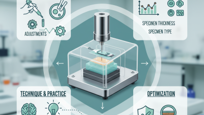
A vibrating microtome cuts with a side-to-side blade motion, helping reduce compression in soft, unfixed tissue. Vibration alone does not guarantee a clean section. Blade speed, oscillation amplitude, tissue temperature, and specimen support all interact. If the blade advances too quickly, tissue may bunch ahead of its edge. Slow the feed, and cutting time increases, while delicate tissue can still tear.

Current Protocols in Neuroscience describes acute brain slices commonly prepared at roughly 300–400 μm for electrophysiology. That practical range shows why thickness and tissue purpose matter; it is not a universal setting. Higher amplitude may help the blade pass through tougher tissue, but excessive movement can leave rippled edges. Small changes matter. A useful starting point is a firm specimen bed, chilled buffer when appropriate, and gradual adjustments to one setting at a time. Record each result under consistent lighting.

In routine lab work, inspect the section under a low-power microscope before changing several controls at once. Look for compressed corners, chatter marks, and uneven thickness. These clues are more useful than relying on the dial alone. Blade sharpness and mounting quality can confound a trial, too. Not every cut improves. That is worth admitting: optimizing one specimen may not transfer neatly to another, especially when fixation and tissue density differ.

Key Applications: Fresh-Tissue Slices for Neuroscience and Histology

Fresh-tissue slices let neuroscientists examine circuits while many cells remain viable. A vibrating microtome moves its blade through immersed tissue with limited compression, helping preserve delicate layers in brain samples. Slice thickness depends on the experiment: acute electrophysiology often uses sections a few hundred micrometres thick, while thinner sections can suit imaging and staining. Keep the sample cool and oxygenated. Small temperature or vibration changes can still affect tissue quality.

The scale of modern neuroscience makes reproducible preparation essential. The BRAIN Initiative Cell Census Network’s cross-species motor-cortex atlas, published in Nature, analyzed more than 1.3 million cells, underscoring the value of consistent tissue sampling and analysis. For histology, fresh slices support rapid fixation or targeted staining, allowing researchers to compare regions before processing changes obscure fine structures. A clean cut matters. So does orientation: a slight tilt can distort anatomical boundaries and complicate comparisons.

When evaluating vibrating microtome suppliers, laboratories should check blade control, bath temperature stability, and repeatable thickness settings. Ask for demonstrations using tissue similar to the intended specimen; a smooth cut in soft test material is not proof of performance on fragile neural tissue. These checks are easy to overlook. Even a well-adjusted instrument cannot rescue tissue that was poorly handled, and slice quality should be verified under a microscope rather than assumed from settings alone.

Typical Vibrating Microtome Slice Thickness by Application

Indicative working ranges for fresh-tissue sectioning: thicker slices are commonly used for electrophysiology, while thinner sections are often used for free-floating histology. Actual thickness depends on tissue, species, and experimental protocol; these ranges are not universal specifications.

How to Compare Chinese Manufacturers: Specifications, Support, and Compliance

When comparing Chinese vibrating microtome manufacturers, request complete specifications, not just maximum cutting speed. Check section thickness range, vibration frequency, blade-angle adjustment, specimen capacity, and compatibility with your sample types. Ask suppliers to demonstrate a cut on a similar tissue block. Small details matter. A stable setup should produce consistent sections without excessive compression or tearing.

The ISO Survey of Management System Standard Certifications 2022 recorded about 1.27 million valid ISO 9001 certificates worldwide. This figure shows how widely quality systems are used, but certification alone does not prove a microtome’s performance. Request the certificate’s scope and verify that it covers the manufacturing site. Also ask for inspection records, calibration procedures, electrical safety documentation, and clear instructions for cleaning and maintenance. Paperwork matters.

Support can determine how quickly a lab returns to work after a fault. Ask about spare-blade availability, remote troubleshooting, training, warranty terms, and realistic response times for your location. A live demonstration is useful, but one successful cut is not enough; request repeat tests and inspect section consistency under a microscope. I would also compare the full service offer, not just the purchase price. Some specifications look impressive on paper, yet routine usability can be less obvious.

Supplier Due Diligence: Verify Performance Data, Quality Systems, and Service Coverage

When assessing China-based vibrating microtome manufacturers and suppliers, request performance data tied to clear test conditions. Ask about section thickness range, cutting speed, vibration frequency, and repeatability across comparable tissue samples. A number without a method is hard to judge. Request sample records or a live demonstration, and check whether the tested settings match your intended workload. Watch how the instrument handles a delicate sample, not just an easy demonstration specimen. Results can vary with tissue preparation and operator technique, so avoid treating one successful cut as proof of consistent performance.

Review the quality system through controlled documents, calibration records, component traceability, and procedures for handling defects. Confirm which checks are performed before shipment and whether inspection results can be linked to the unit’s serial number. Then verify service coverage in writing: installation support, operator training, spare-part availability, response times, and the process for resolving faults. Ask who handles service in your region and what happens if a key part is unavailable. A polished brochure is not evidence. Supplier explanations may sound complete, yet missing records deserve follow-up. It is easy to overlook routine details, such as blade holders or replacement seals; include them in the review.

preloader